


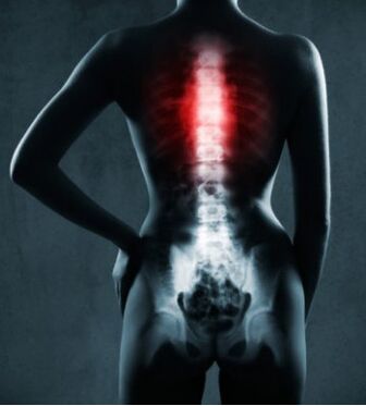
Thoracic osteochondrosis is a degenerative lesion of the spine (exhaustion and destruction of the bony structure of the vertebrae). It begins with a behavioral wound, the onset of autonomic symptoms (shortness of breath, weakness, sweating, malaise) and the development of severe pain syndrome. Thoracic osteochondrosis mimics cardiovascular disease and therefore requires an accurate differential diagnosis. Therapy includes a wide range of treatments: medication, exercise therapy, physiotherapy and massage.
Thoracic osteochondrosis is less common than the cervix or lumbar spine. This is due to the specifics of the anatomical structure. The vertebral discs in the thoracic region occupy two-thirds of the entire spine in number, and they are also larger in diameter but inferior in size to the lumbar region. This area is strong and has low mobility, and is protected by ribs and ribs. The physiological curvature is directed towards the back. This leads to increased stress on the front of the spine. Further, the formation and growth of pathological bone structures in the vertebral bodies (osteophytes) occurs. Peripheral nerve endings are located between ligaments and muscle tissue, their tension leads to compression with the development of pain.
There are also polysgmented spinal lesions with osteochondrosis. At the same time, degeneration of the cervical, thoracic, and lumbar regions is combined with the corresponding clinical symptoms.
The clinical symptoms of thoracic osteochondrosis among women and men are approximately the same and have no significant differences.
Prevalence
Diagnosis can be made at any age. The disease is common in adolescents with a weak musculoskeletal system, as well as as a result of their active growth. The pathology often forms among pregnant women due to the pronounced load on the chest region during pregnancy.
Everyone has a predisposition to the formation of osteochondrosis in the chest region. This is due to the upright posture of a person and, as a result, of a large load on the spinal section.
Distribution
Thoracic pain syndrome is characterized by severe chest pain of an intense nature. The syndrome is associated with peripheral nerve damage. The loss is due to compression of the nerves by the muscles and ligaments.
Degrees of thoracic osteochondrosis:
- The first stage is characterized by the absence of pronounced clinical manifestations. There is a loss of elasticity from the intervertebral discs, and their extensions are formed.
- The second degree is marked by a further loss of elasticity of the intervertebral discs and a decrease in their height. The likelihood of a hernia increases. Pain syndrome appears, symptoms of accompanying pain are possible.
- In the third degree, the pain syndrome increases. The appearance of a herniated disc located between the vertebrae is possible. The severity of the symptoms depends on the location of the hernia.
- Fourth degree with a complete violation of the elasticity and loss of function of the intervertebral discs, destruction of the bone structure of the vertebrae. Neurological disorders are more pronounced.
According to the types of pain symptoms:
- Spinal thoracalgia is justified by spinal pathology.
- Non-vertebrogenic thoracalgia is caused by the formation of pathologies of internal organs: cardiovascular diseases, gastroduodenal reflux, traumatic and inflammatory lesions of the musculoskeletal system.
- Psychogenic thoracalgia is caused by panic attacks and damage to the organs of nervous genesis.
Causes and risk factors
Osteochondrosis does not form without lesion factors. A number of reasons or a combination of them leads to the development of the disease in the chest region.
- Sedentary lifestyle. Lack of physical activity leads to weakness of the muscles of the back and intervertebral segment. Sedentary work and improper workplace organization acts as an additional factor in thoracic osteochondrosis.
- Improper weight lifting and a range of injuries. Excessive stress that disrupts the functioning of the spine. In this situation, the muscles and intervertebral discs cannot withstand the load.
- Acquired lesions and curvature of the spine. Against the background of these pathologies, the work of the spine is interrupted and the likelihood of the formation of osteochondrosis increases. The destruction intensifies if the doctor's recommendations are not followed.
- Lack of required minerals and vitamins. With an insufficient concentration of calcium in the bone tissue, the bones become weaker and the likelihood of damage to the musculoskeletal system increases.
- Pregnancy as a combination of key factors: an increased load on the spine and a lack of minerals and vitamins.
Important!Hereditary predisposition plays an important role. If lesions of the musculoskeletal system are observed along a connected line, then you should take care of your health and prevent lesions. A competent system of preventive measures prevents the massive destruction of bone tissue.
Who is at risk
Often the factors for the formation of degenerative changes in the spine are combined.
- A decrease in immune status accompanied by a greater susceptibility to infections that may increase the clinical manifestations of osteochondrosis due to muscle inflammation.
- Stressful effects that can cause psychogenic thoracalgia. This is due to the large release of catecholamines, which provoke an increase in pain.
- Nervous system damage of non-infectious and infectious etiology.
- Physical overload.
- Non-compliance with the principles of ergonomics (carrying weights).
- Spinal cord injuries of various origins.
- Muscle spasms.
- Osteoporotic degeneration of the musculoskeletal system.
Symptoms
The main symptoms of thoracic osteochondrosis
- The burning sensation that arises in the intercostal spaces.
- Paroxysmal and persistent chest pain, mainly knives.
- With thoracalgia, the pain syndrome is stabbing, tightening and painful.
- Pain in the groin.
- Pain on one side of the trunk.
- During movement, a vertebral crisis is observed.
- Pain symptoms increase significantly with movement, deep breathing, coughing and sneezing, which is the main difference between thoracic osteochondrosis and angina pectoris.
- The affected areas are vulnerable, i. e. they can be felt and located along the affected nerves.
- Numbness of the skin along the intercostal spaces.
- The patient's condition worsens when exposed to low temperatures or prolonged stay in an uncomfortable position.
Varieties of pain syndromes in osteochondrosis of the thoracic spine:
- Lower neck lesion. There is soreness in the upper chest, which can radiate to the neck, arms, and also to the left half of the body.
- Injury of the upper thoracic spine. The pain is painful in nature, affects the central part of the chest. Frequent combination with pain in the shoulder blade area.
- Loss of scapular-coastal area. Painful symptoms have a sharp, painful and stinging character. It has the appearance of attacks, long and short. It occupies the lateral region, and also focuses on the shoulder blade area.
- The appearance of pain in the front wall of the chest, which varies in duration. They arise between the peri-pectoral and frontal axillary lines.
In addition to the main signs, there are two types of pain syndromes in thoracic osteochondrosis:
- Dorsago - severe but short-term pain at the site of localization of the affected intervertebral discs. Disturbance of normal breathing.
- Dorsalgia - mild but prolonged pain in the area of the affected intervertebral discs.
Spondylogenetic thoracalgiaaccompanied by damage to the musculoskeletal system, is often accompanied by severe pain and instability of the vertebrae in the thoracic spine (their increased mobility). The loss is expressed in a violation of the mobility of the thoracic spine, suturing and cutting pain in the intercostal spaces.
Vertebral thoracalgiamay provoke the following symptoms:
- radicular (pain symptoms);
- violation of thoracic area innervation (manifestations of internal organs: a number of patients have painful symptoms of a stabbing nature in the digestive tract or cardiovascular system);
- radicular syndrome with vegetative signs (soreness in the intercostal spaces).
When diagnosing a problem, you are required to distinguish symptoms from cardiovascular disease and myalgia. Heart damage of ischemic etiology is distinguished by the regularity of occurrence during physical or psycho-emotional stress and the relief of an attack by taking nitrates.
A psychogenic attack of thoracic pain is associated with the onset of panic, anxiety, choking, and mental disorder. It turns out that the disease is a consequence of problems with psychological stability.
The clinical signs of osteochondrosis are divided into two main parts:
- Neuralgic symptoms:
- With thoracic osteochondrosis, numbness and tingling can occur both in the upper limbs and along the intercostal spaces, spreading to the anterior surface of the chest.
- The latissimus dorsi and chest muscles are in constant tension.
- There is a high emotional calm, periods of tears and nervousness.
- In rare situations, the disease presents as pronounced intercostal neuralgia.
- Different types of pain sensations:
- Dorsago: sharp, acute soreness in the thoracic spine, sometimes making breathing difficult. Movement in the spine, cervix and thorax is limited. Manifested or worsens when sitting in a twisted position.
- Dorsalgia: the formation of pain symptoms lasts from two to three weeks, therefore, at first it continues without clinical manifestations for the patient. There is a slight discomfort in the chest. The pain is aggravated by turning the body sideways and taking deep breaths. With the final stabilization of the pathological process, a persistent pain syndrome is formed.
- Intercostal neuralgia: band pain that radiates along the intercostal spaces. When you breathe sharply, a stabbing pain appears in the region of the heart. As a result, the pathology is often confused with damage to the cardiovascular system.
- Cardiac or pseudocoronary syndrome is formed by lesions at the level of the ThI segments with the development of reflex angina pectoris. The difference from organ damage to the cardiovascular system is the appearance of pain when you bend or rotate your spine. They intensify with prolonged stay in a forced position. There is pain on palpation of spinous processes in the thoracic spine.
- Radicular syndrome: soreness in the intercostal spaces (Erb points).
- Internal organ syndrome: dysfunction of the abdominal organs with lesions at the level of thoracic vertebrae V-XII. Expressed in girdle pain, aggravation in proper hypochondrium, heartburn.
Clinical symptoms depending on the level of thoracic spine lesion:
* Loss of nerve processes in thoracic osteochondrosis occurs in cases of the appearance of osteophytes - bone protrusions in the vertebrae. This is due to the degree of destruction. Therefore, the symptoms below are not an integral part of the disease.
- Deformation of the nervous process at Th2 and Th3 levels. Damage to the cardiovascular system occurs with the onset of arrhythmia attacks and coronary heart disease. Thus, the symptoms of chronic pain in thoracalgia can provoke dysfunction of the organs of the cardiovascular system.
- Loss at Th4-Th5 level. Organs with damaged nerve fibers: pleura and bronchitis, pneumonia, bronchial asthma.
- Th5-Th6: bile ducts and gallbladder affected. The absorption of fats in the body is reduced.
- Th6-Th7: affects the liver and the solar plexus area. The function of the hepatobiliary tract is impaired.
- Th7-Th8: stomach affected. Main pathologies: ulcerative lesions of the duodenum and stomach, indigestion and gastritis.
- Th8-Th9: changes in the functioning of the duodenum and pancreas. Manifestations: duodenitis, pancreatitis and loose stools.
- Th9-Th10: damage to nerve cells of internal organs (spleen and diaphragm). There are hiccups and breathing problems.
- Th10-Th11: adrenal glands are affected. The activity of the immune system decreases and allergies appear.
- Th11-Th12: kidney function is impaired, leading to the formation of pyelonephritis and urolithiasis.
- Th12-L1 (level of the first lumbar vertebra). The kidneys and ureters are damaged. This leads to dizziness - problems with urination.
Diagnosis of thoracic osteochondrosis
If you suspect osteochondrosis, you can contact a therapist or neurologist.
The patient is examined with the recording of all clinical data. During the formation of stages 2-3, the skeleton undergoes significant deformation. A complete history of the patient should be compiled in order to accurately determine or rule out the factors that lead to the formation of osteochondrosis of the thoracic spine.
The first diagnostic method is radiography. Further studies are performed based on clinical history data and the need for differential diagnosis.
Any doctor can first examine the patient. The main thing is a competent and fully collected clinical history. This will allow you to accurately determine the etiology of the disease and choose a therapy regimen. Therapist, neurologist, rheumatologist are involved in the treatment of thoracic osteochondrosis. In case of traumatic effects on the spinal region, a consultation with a traumatologist is required.
- X-ray examination of the chest in two projections. Allows you to determine the presence and size of osteophytes, determine the contours and height of intervertebral discs, make changes in the shape of the disc.
- Discography makes it possible to examine the structure of the pulposus nucleus through the use of contrast.
- Computed tomography is used to visualize nerve fibers, muscles, ligaments, and joints.
- Electromyography allows differential diagnosis with neurological diseases.
- Endoscopic diagnostic methods can be prescribed for the purpose of examining the circulatory and digestive organs.
- An ECG is performed to determine the etiology of cardiovascular disease.
- Electroencephalography - to determine the pathologies of the nervous system.
Differential diagnosis
Thoracic osteochondrosis must be distinguished from a number of diseases.
- Anomaly in the formation of the spine, trauma, tumor, inflammation. There are several possibilities for these pathologies. For example, an additional congenital process, displacement or fusion of vertebrae (spondylolisthesis), osteomyelitis, ankylosing spondylitis and others.
- Damage to the musculoskeletal system (various lengths of the lower limbs, muscle spasms, muscle inflammation and others).
- It is not associated with damage to the musculoskeletal system, but similar in symptoms of diseases of internal organs. In particular, pancreatitis, inflammation of the appendages, stomach ulcers, coronary heart disease, angina pectoris, pleurisy.
- Neurosis-like disorders, combined with migratory pain with increased fatigue, irritability, mood swings.
Chest osteochondrosis and ischemic heart disease
It is extremely important to carry out a competent differential diagnosis with the most similar pathologies. Pain arising from vertebral thoracalgia and coronary heart disease (FDI) have a number of variations, which make it possible to make an accurate diagnosis.
Nature of pain: with coronary artery disease, they have a burning and constricting character, accompanied by fear of death.
From the duration of the pain:
- FDI: Short-term, attack within minutes.
- Chest osteochondrosis is characterized by mild or prolonged pain, in some cases they do not subside during the day.
Change in body position:
- With ischemic heart disease, the strength and intensity of pain do not change with physical activity.
- With thoracalgia, even relatively light movements cause increased pain or the appearance of a new attack.
Reaction to physical activity:
- With ischemic heart disease, the pain appears during physical exertion, stopping at rest.
- Thoracalgia, on the other hand, weakens but does not stop at rest.
Medication fillers:
- With an ischemic attack, the pain is easily relieved by taking nitrates.
- Thoracalgia is relieved by the use of analgesics.
Influence of physiotherapeutic factors and manual therapy:
- With ischemic heart disease, it gives an unstable and easy improvement.
- With osteochondrosis, there is a significant positive dynamic in the patient's condition.
Treatment of chest osteochondrosis
Osteochondrosis is treated by a neurologist.
For the organization of competent therapy, it is first required to determine the etiological preconditions. Identifying the cause of the pathology allows you to choose the right treatment regimen.
Preparations for bone tissue regeneration are selected taking into account all the functional characteristics of the body. It is advisable to clarify in advance the concentration of collagen and elastin in the body. When choosing a therapy regimen, the individual characteristics of the organism are taken into account.
Standard therapy regimen
Non-steroidal anti-inflammatory drugs help relieve chest pain caused by inflammatory reactions. This increases the volume of chest mobility as well as the range of motion in the spine.
Drugs that affect the production of interleukins. They allow to stop the inflammatory cascade and normalize the balance of enzymes that cause the destruction of myelin sheaths.
Antispasmodics are also used.
B vitamins help stop the inflammation of the affected nerves.
Preparations that maintain the concentration of collagen and elastin allow you to retain fluid in the intervertebral discs. This increases tissue elasticity and prevents further degeneration.
Hormonal drugs (steroids). They have a powerful anti-inflammatory effect, but are used only for acute thoracalgia, as they negatively affect the body as a whole.
Diuretics in the acute period of the disease help relieve swelling from nerve endings. Preference is given to potassium-sparing diuretics.
Ointments and anti-inflammatory gels. When you rub the affected area of the back, the local inflammatory process is reduced and the symptoms of very active pain are eliminated.
Massage
The therapeutic effect of massage is the relief of spasm from the muscular corset of the thoracic spine and the normalization of local blood circulation.
Effects of massage techniques:
- removal of muscular hypertension;
- strengthening the structure of intervertebral disc bodies.
The use of massage techniques is combined with a visit to a chiropractor in combination with a regular system of exercise therapy.
Physiotherapy
Acupuncture. Eliminates or reduces muscle spasm, and also reduces pain symptoms.
Manual therapy. Allows you to bring the systemic circulation to a normal state in the intercostal space. This conditions the supply of nutrients to the tissues, improves their trophism and stimulates blood oxygenation.
Diet for osteochondrosis of the thoracic spine
Compliance with certain nutritional principles allows you to achieve the maximum therapeutic effect.
- Foods rich in vitamins A, B, C and E (greens, nuts, whole grains) are recommended.
- Omega-3 fatty acids. 6 found in fish.
- Cartilage tissue regeneration stimulants in the form of food additives allow the maintenance of tissue strength and maintain the elasticity of tissue structures.
Complications
When establishing the diagnosis of thoracic osteochondrosis, the possible cascade of possible organ pathologies that develop over time should be considered.
- Damage to the cardiovascular system: persistent pain syndrome leads to destabilization of ion exchange of the myocardial muscle, which is a prerequisite for the development of coronary heart disease.
- Disorders of the functioning of the abdominal organs: stomach, duodenum, pancreas. This is due to the high secretion of adrenaline with persistent pain syndrome, which leads to increased secretion of VIP (vasointestinal peptides).
- Gallbladder dyskinesia is justified by an increase in biliary lithogenicity against the background of a chronic inflammatory process.
With regular adherence to the principles of therapy, exercise therapy system, behavioral maintenance and elimination of risk factors, the course of the disease is reduced in regression. The prognosis is considered favorable if the pathology does not develop further and the disease does not appear actively.
Prophylaxis
- Elimination of hypodynamics, therapeutic exercises. Exercises against force, perpendicular loads with displacement, spine extension are chosen.
- When driving a car for a long time, the choice of special exercises to relax the muscle frame.
- Pumping the muscles of the thoracic spine. There is a complex of exercise therapy and the use of myostimulation when independent training is impossible.
- Workplace organization: the back of the work chair should provide support for the spine. So that the load on the spine does not increase, you need to warm up every 30 minutes in the form of lying down or walking. This is because the sitting position puts more stress on the back.
- Accurate spine position at night: buy orthopedic sleeping equipment. A completely rigid surface is not rational due to the violation of physiological curves of the spine.
- Compliance with ergonomics principles: do not lift weights that could damage the spine.
- Formation of correct behavior.
- Optimization of blood circulation and lymphatic flow through a system of stretch marks or the use of special procedures (typotherapy).